Vomiting in neonate Causes & diagnosis

16 Slides84.68 KB
Vomiting in neonate Causes & diagnosis
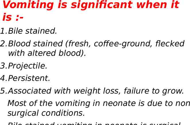
Vomiting is significant when it is :1.Bile stained. 2.Blood stained (fresh, coffee-ground, flecked with altered blood). 3.Projectile. 4.Persistent. 5.Associated with weight loss, failure to grow. Most of the vomiting in neonate is due to non surgical conditions.
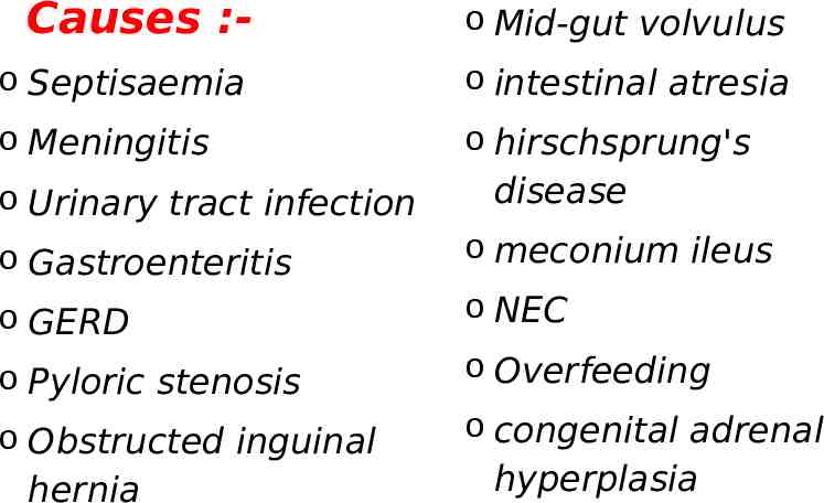
Causes :- o Mid-gut volvulus o Septisaemia o intestinal atresia o Meningitis o hirschsprung's o Urinary tract infection disease o Gastroenteritis o meconium ileus o GERD o NEC o Pyloric stenosis o Overfeeding o Obstructed inguinal hernia o congenital adrenal hyperplasia
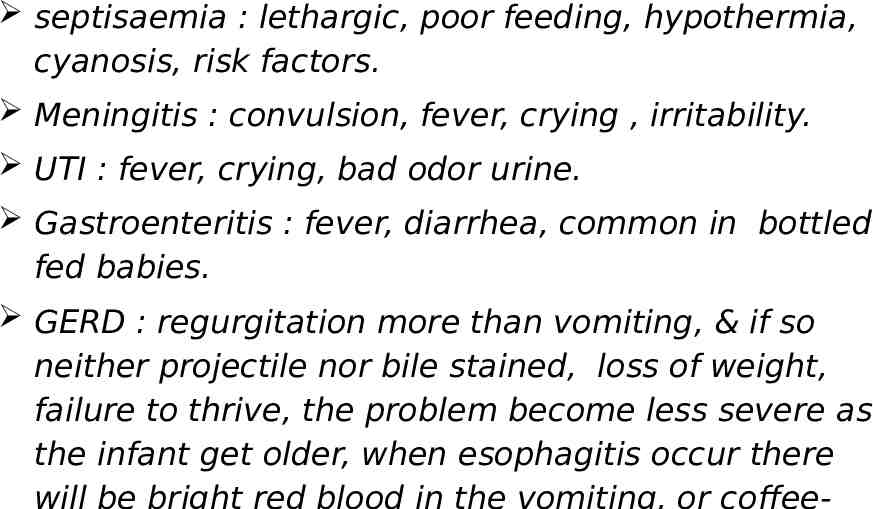
septisaemia : lethargic, poor feeding, hypothermia, cyanosis, risk factors. Meningitis : convulsion, fever, crying , irritability. UTI : fever, crying, bad odor urine. Gastroenteritis : fever, diarrhea, common in bottled fed babies. GERD : regurgitation more than vomiting, & if so neither projectile nor bile stained, loss of weight, failure to thrive, the problem become less severe as the infant get older, when esophagitis occur there
Pyloric stenosis : The most common surgical cause of vomiting in infant, M : F 4 : 1, Non bile stained vomiting, projectile, eager to feed following vomiting, on examination; visible gastric peristalsis , palpable olive mass which confirm the diagnosis (felt in the epigastrium or just to the right of the rectus abdominus musle. Failure to palpate the mass :o Be patient, re-examine when he is quite or asleep. o Be gentle o Flex the hip joint
Differential diagnosis of the mass:1. Pole of right kidney 2. Caudate lobe of the liver 3. Tip of NG tube 4. Body of vertebrae Visible gastric peristalsis & projectile vomiting are supportive but not by themselves diagnostic. Ultrasound : length of pyloric canal & thickness if 16 mm, 4mm. Barium meal : string sign (long narrow pyloric canal) Shoulder sign (impression of pylorus into the duodenal mucosa)
Obstructed inguinal hernia :Nearly all inguinal hernia in infant is (indirect). M:F 8:1 . 60 % in the right side, 25% in the left side, 15 % bilateral. The most common condition requiring surgery in childhood. Incidence 1-2 per 100 live births male. 30% firstly presented with strangulated hernia. Presentation of inguinal hernia : history of intermittent swelling overlying external inguinal ring painless or with occasional discomfort, bulge on crying or straining. Silk glove sign : contiguous layer of peritoneum of empty sac. Thickened spermatic cord in comparism to the other side. Presentation of obstructed inguinal hernia : crying , vomiting, abdominal distention, irreducible swelling in the groin which is tens & tender. With the delay in the diagnosis there will be induration overlying the lump, redness, hotness which are signs of peritonitis & bowel ischemia.
Differential diagnosis :o Encysted hydrocele of the cord o Undescended testis (empty scrotal sac on the affected side) o Torsion of the testis o Lymphadenitis or local inguinal abscess.
Volvulus neonatorum :In Malrotation of midgut there is narrow mesentery of midgut & labile to twist around the axis of superior mesenteric artery. It is emergency condition requiring urgent intervention. Presentation : healthy full term baby who is well for the first few days of life the suddenly developing bile stained vomiting then abdominal distention, blood per rectum, peritonitis, septisaemia.
Intestinal atresia :o o o o Pyloric atresia Duodenal atresia Jejunoileal atresia Colonic atresia Antenatal ultrasound show polyhydramnios Down syndrome is common associated anomaly Symptoms : vomiting , abdominal distention , constipation. Diagnosis : Plain x-ray : single bubble Double bubble Air fluid levels Gasless lower abdomen Contrast study (barium enema) : microcolon.
Hirschsprung's disease :Delayed or non passing meconium Abdominal distention Bile stained vomiting Rectal examination with probe : explosive decompression of meconium & feces Investigations : Contrast enema : narrow, transitional ,& dilated segments. Rectal biopsy : absent ganglion cells, hypertrophic nerve fibers, increase staining with cholin esterase.
NEC(necrotizing enterocolitis) :95% occur in pre mature. Risk factors : Presentation : lethargic, poor feeding, abdominal distention, bile stained vomiting , blood per rectum, then progress to peritonitis, edematous redness of the anterior abdominal wall, palpable mass of intra abdominal abscess. Diagnosis : Plain x- ray : air fluid levels Pneumatosis intestinalis Portal venous gas Air under diaphragm
Meconium ileus :15% 0f cystic fibrosis firstly presented with meconium ileus. Genetic mutation ΔF508 in the cell membrane protein CFTR. Cystic fibrosis causes changes in the composition of meconium (thicker, sticky,& tenacious). Presentation : bile stained vomiting, abdominal distention, failure to pass meconium, loops of distended gut palpable as they filled with meconium rather than gaseous distention. Rectal examination : no normal meconium, there is pellets of mucus. Plain x-ray : no air fluid level, ground glass appearance, soap bubble appearance. Contrast study : microcolon with pellets in the terminal ileum.
Congenital adrenal hyperplasia :21- hydroxylase deficiency will cause block in the synthesis of cortisol & aldosterone & 17-hydroxyprogesterone shift to synthesis of androgen. Increase stimulation of ACTH : adrenal hyperplasia Decrease cortisol : hypoglycemia Decrease aldosterone : salt losing metabolic disturbance (vomiting) which life threatening situation. Increase androgen : virilization of female body & ambiguous genitalia.
Overfeeding :In bottle fed babies Healthy ( no weight loss) Improper feeding habits. Dr.Ali E. Joda