PULSE K. JAI SHANKAR MD,DM CONSULTANT CARDIOLOGIST INSTITUTE


84 Slides1.33 MB
PULSE K. JAI SHANKAR MD,DM CONSULTANT CARDIOLOGIST INSTITUTE OF CARDIOVASCULAR DISEASES MADRAS MEDICAL MISSION
PULSE DEFINITION: Pulse is the palpability over peripheral arteries, a pulse wave which is a transmitted wave from the root of aorta along the vessel wall traveling 10 times faster than blood. Blood travels at speed of - .5 mt/sec. Pulse travels at speed of - 5 mt/sec.
PULSE WAVE The arterial pulse reflects the performance of LV “Mirror of the heart” It is propagated by incompressible blood both forwards and laterally. The lateral movement distends the arterial wall and is felt as pulse.
PULSE - HISTORY HIPPOCRATES – 4TH CENTURY BC Thought that arteries are air ducts GALEN Arteries contain blood & not air. HEROPHILUS Recognized that arterial pulses & cardiac pulses were synchronous.
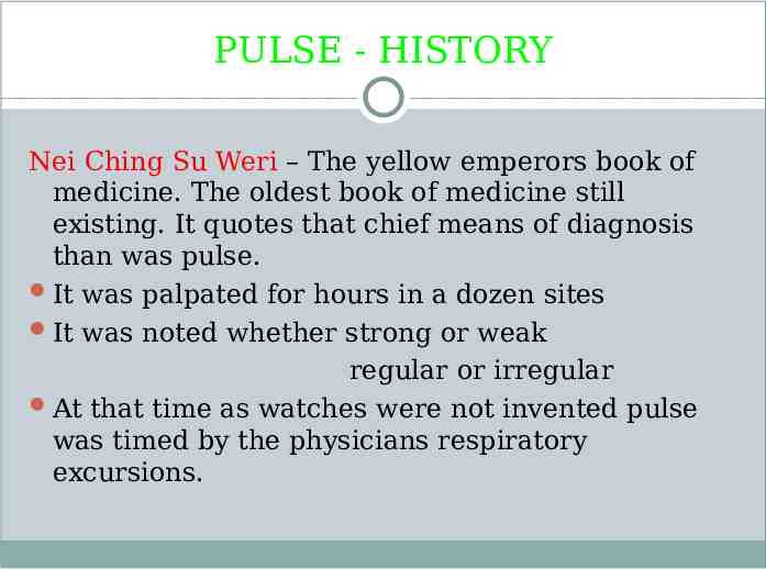
PULSE - HISTORY Nei Ching Su Weri – The yellow emperors book of medicine. The oldest book of medicine still existing. It quotes that chief means of diagnosis than was pulse. It was palpated for hours in a dozen sites It was noted whether strong or weak regular or irregular At that time as watches were not invented pulse was timed by the physicians respiratory excursions.
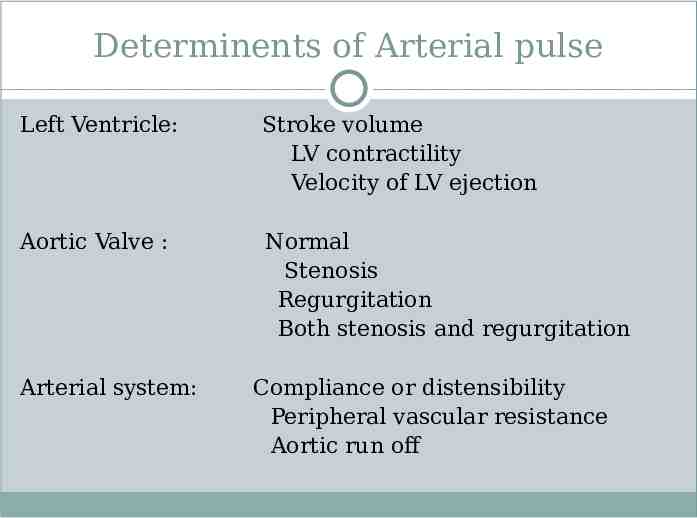
Determinents of Arterial pulse Left Ventricle: Stroke volume LV contractility Velocity of LV ejection Aortic Valve : Normal Stenosis Regurgitation Both stenosis and regurgitation Arterial system: Compliance or distensibility Peripheral vascular resistance Aortic run off
BLOOD FLOW LV pressure when it rises above aortic pressure becomes driving force for movement of blood into aorta Driving force is dependent on 1) Contractility 2) Size & shape of LV 3) Heart rate. This driving force is opposed by several forces that impede the flow 1) Resistance2) Inertia3) Compliance
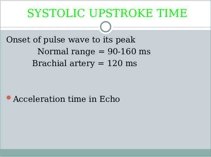
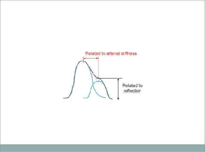
SYSTOLIC UPSTROKE TIME Onset of pulse wave to its peak Normal range 90-160 ms Brachial artery 120 ms Acceleration time in Echo
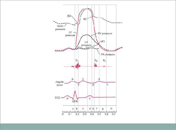
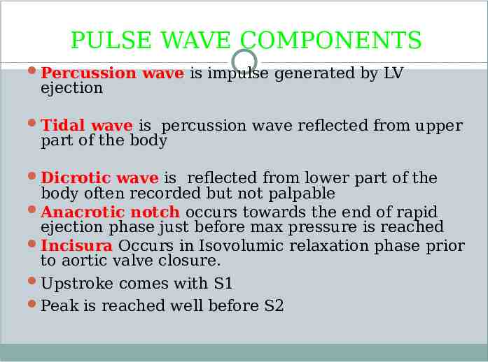
PULSE WAVE COMPONENTS Percussion wave is impulse generated by LV ejection Tidal wave is percussion wave reflected from upper part of the body Dicrotic wave is reflected from lower part of the body often recorded but not palpable Anacrotic notch occurs towards the end of rapid ejection phase just before max pressure is reached Incisura Occurs in Isovolumic relaxation phase prior to aortic valve closure. Upstroke comes with S1 Peak is reached well before S2
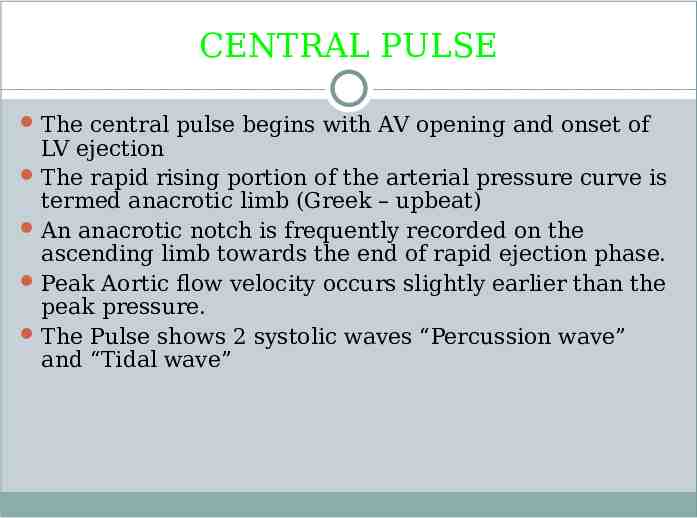
CENTRAL PULSE The central pulse begins with AV opening and onset of LV ejection The rapid rising portion of the arterial pressure curve is termed anacrotic limb (Greek – upbeat) An anacrotic notch is frequently recorded on the ascending limb towards the end of rapid ejection phase. Peak Aortic flow velocity occurs slightly earlier than the peak pressure. The Pulse shows 2 systolic waves “Percussion wave” and “Tidal wave”
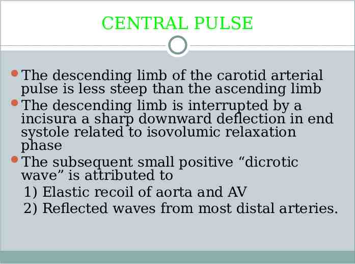
CENTRAL PULSE The descending limb of the carotid arterial pulse is less steep than the ascending limb The descending limb is interrupted by a incisura a sharp downward deflection in end systole related to isovolumic relaxation phase The subsequent small positive “dicrotic wave” is attributed to 1) Elastic recoil of aorta and AV 2) Reflected waves from most distal arteries.
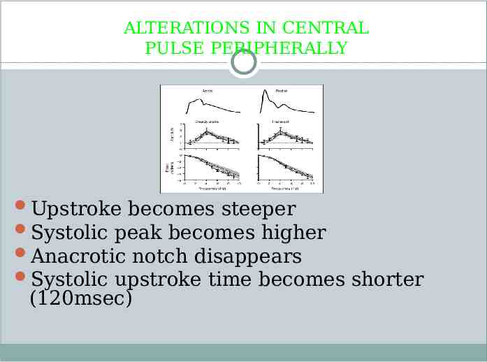
ALTERATIONS IN CENTRAL PULSE PERIPHERALLY Upstroke becomes steeper Systolic peak becomes higher Anacrotic notch disappears Systolic upstroke time becomes shorter (120msec)
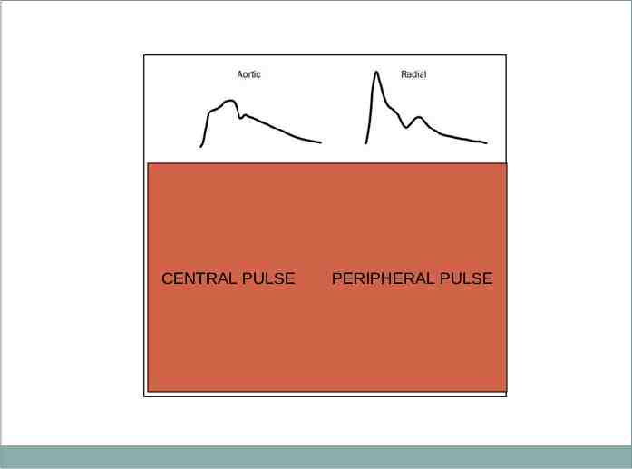
CENTRAL PULSE PERIPHERAL PULSE
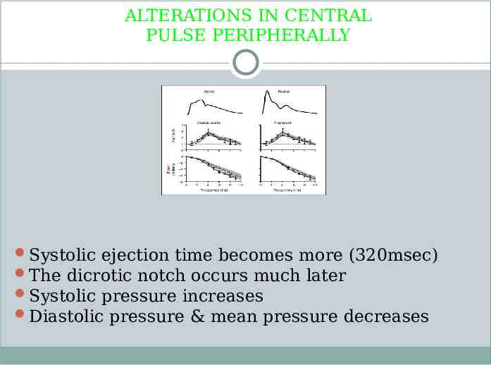
ALTERATIONS IN CENTRAL PULSE PERIPHERALLY Systolic ejection time becomes more (320msec) The dicrotic notch occurs much later Systolic pressure increases Diastolic pressure & mean pressure decreases
CAUSES FOR CHANGE IN CENTRAL PULSE CONTOUR WHEN TRANSMITTED PERIPHERALLY 1)Distortion & damping of pulse wave components 2) Different rates of transmission of various components 3) Differences in distensibility & caliber of arteries 4) Changes in the vessel wall due to age & or disease
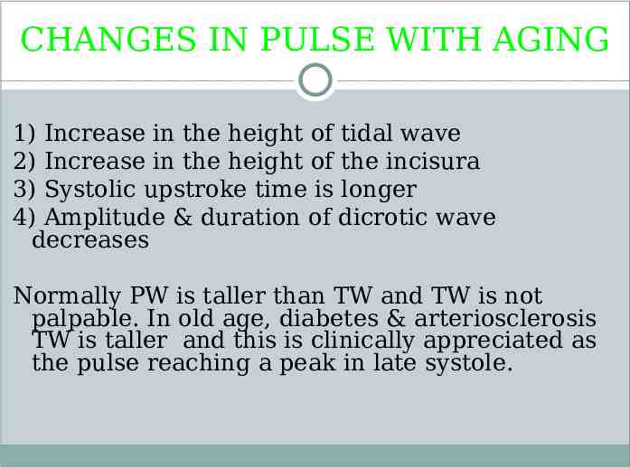
CHANGES IN PULSE WITH AGING 1) Increase in the height of tidal wave 2) Increase in the height of the incisura 3) Systolic upstroke time is longer 4) Amplitude & duration of dicrotic wave decreases Normally PW is taller than TW and TW is not palpable. In old age, diabetes & arteriosclerosis TW is taller and this is clinically appreciated as the pulse reaching a peak in late systole.
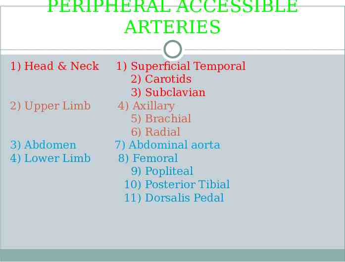
PERIPHERAL ACCESSIBLE ARTERIES 1) Head & Neck 2) Upper Limb 3) Abdomen 4) Lower Limb 1) Superficial Temporal 2) Carotids 3) Subclavian 4) Axillary 5) Brachial 6) Radial 7) Abdominal aorta 8) Femoral 9) Popliteal 10) Posterior Tibial 11) Dorsalis Pedal
Localization of arteries The CCA terminates at C4 level at upper border of thyroid cartilage The ECA is palpated medial to the sternocleidomastoid above upper border of the thyroid cartilage The ICA is palpated placing a hand in the mouth and palpating the tonsillar fauces. The subclavian artery is felt in the posterior triangle. With the shoulder depressed, pressure is exerted down back and medially in the angle between sternocleidomastoid and clavicle.
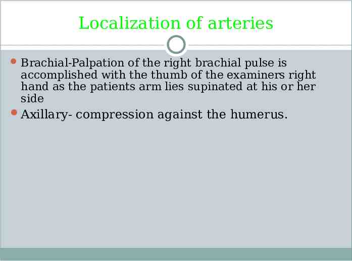
Localization of arteries Brachial-Palpation of the right brachial pulse is accomplished with the thumb of the examiners right hand as the patients arm lies supinated at his or her side Axillary- compression against the humerus.
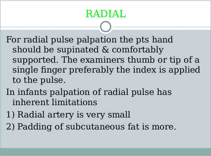
RADIAL For radial pulse palpation the pts hand should be supinated & comfortably supported. The examiners thumb or tip of a single finger preferably the index is applied to the pulse. In infants palpation of radial pulse has inherent limitations 1) Radial artery is very small 2) Padding of subcutaneous fat is more.
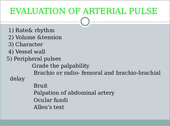
EVALUATION OF ARTERIAL PULSE 1) Rate& rhythm 2) Volume &tension 3) Character 4) Vessel wall 5) Peripheral pulses Grade the palpability Brachio or radio- femoral and brachio-brachial delay Bruit Palpation of abdominal artery Ocular fundi Allen’s test
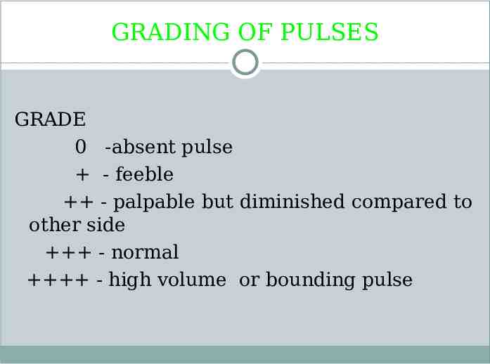
GRADING OF PULSES GRADE 0 -absent pulse - feeble - palpable but diminished compared to other side - normal - high volume or bounding pulse
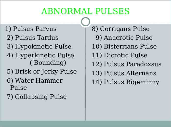
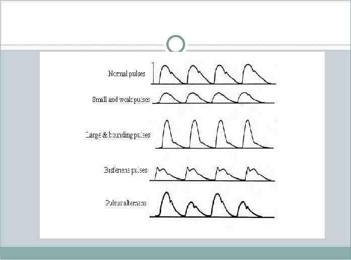
ABNORMAL PULSES 1) Pulsus Parvus 2) Pulsus Tardus 3) Hypokinetic Pulse 4) Hyperkinetic Pulse ( Bounding) 5) Brisk or Jerky Pulse 6) Water Hammer Pulse 7) Collapsing Pulse 8) Corrigans Pulse 9) Anacrotic Pulse 10) Bisferrians Pulse 11) Dicrotic Pulse 12) Pulsus Paradoxsus 13) Pulsus Alternans 14) Pulsus Bigeminny
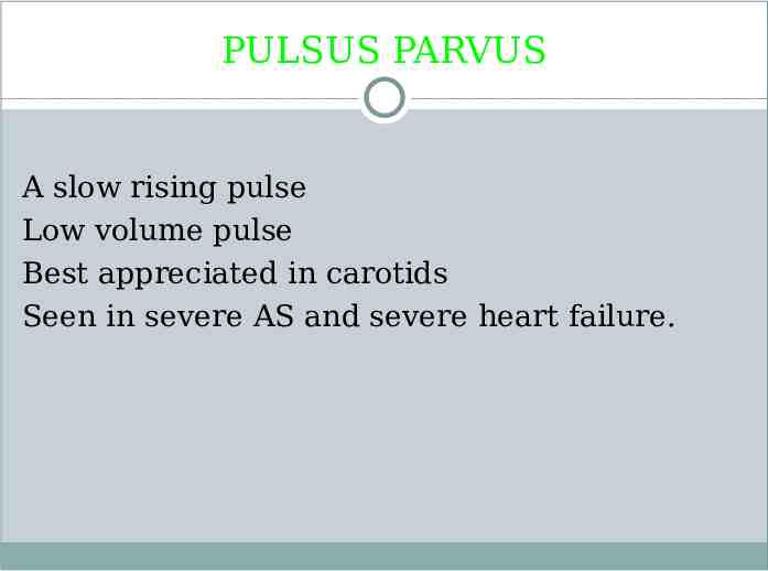
PULSUS PARVUS A slow rising pulse Low volume pulse Best appreciated in carotids Seen in severe AS and severe heart failure.
PULSUS TARDUS( Anacrotic pulse) Late peaking Peak is delayed and nearer to S2 Best appreciated by simultaneous auscultation of the heart and palpation of carotid pulse Seen in all forms of fixed obstruction to the LVOT
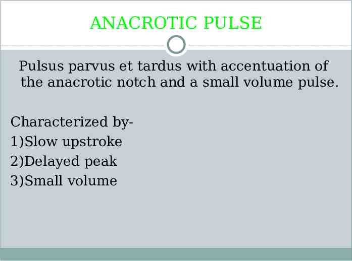
ANACROTIC PULSE Pulsus parvus et tardus with accentuation of the anacrotic notch and a small volume pulse. Characterized by1)Slow upstroke 2)Delayed peak 3)Small volume
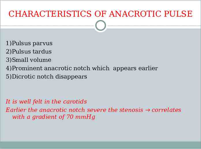
CHARACTERISTICS OF ANACROTIC PULSE 1)Pulsus parvus 2)Pulsus tardus 3)Small volume 4)Prominent anacrotic notch which appears earlier 5)Dicrotic notch disappears It is well felt in the carotids Earlier the anacrotic notch severe the stenosis correlates with a gradient of 70 mmHg
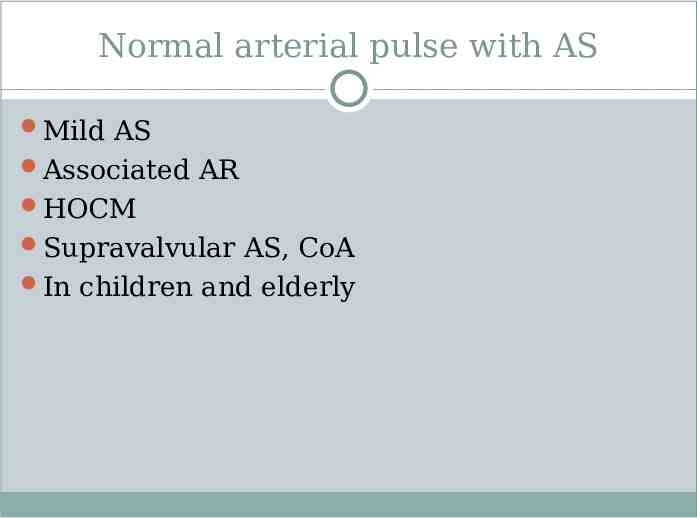
Normal arterial pulse with AS Mild AS Associated AR HOCM Supravalvular AS, CoA In children and elderly
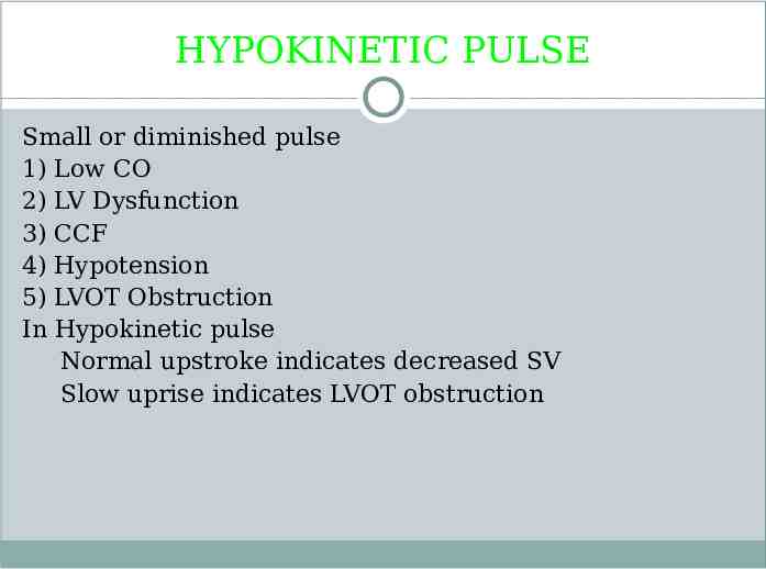
HYPOKINETIC PULSE Small or diminished pulse 1) Low CO 2) LV Dysfunction 3) CCF 4) Hypotension 5) LVOT Obstruction In Hypokinetic pulse Normal upstroke indicates decreased SV Slow uprise indicates LVOT obstruction
HYPERKINETIC PULSE 1) Anxiety 6) Alcohol intake 2) Anaemia 7) Cigarette smoking 3) Thyrotoxicosis 8) SHT with Atherosclerosis 4) Exercise 5) Hot humid environment 9) Isolated Systolic HT
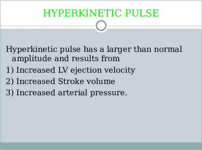
HYPERKINETIC PULSE Hyperkinetic pulse has a larger than normal amplitude and results from 1) Increased LV ejection velocity 2) Increased Stroke volume 3) Increased arterial pressure.
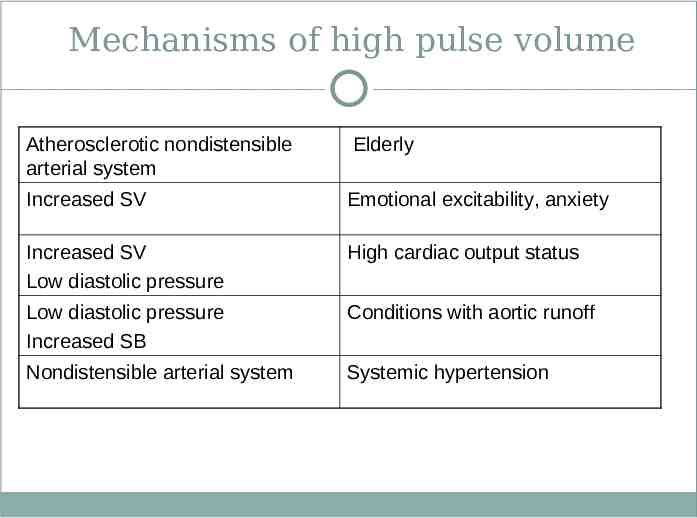
Mechanisms of high pulse volume Atherosclerotic nondistensible arterial system Elderly Increased SV Emotional excitability, anxiety Increased SV Low diastolic pressure High cardiac output status Low diastolic pressure Increased SB Conditions with aortic runoff Nondistensible arterial system Systemic hypertension
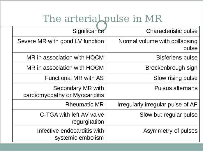
The arterial pulse in MR Significance Characteristic pulse Severe MR with good LV function Normal volume with collapsing pulse MR in association with HOCM Bisferiens pulse MR in association with HOCM Brockenbrough sign Functional MR with AS Slow rising pulse Secondary MR with cardiomyopathy or Myocariditis Pulsus alternans Rheumatic MR Irregularly irregular pulse of AF C-TGA with left AV valve regurgitation Slow but regular pulse Infective endocarditis with systemic embolism Asymmetry of pulses
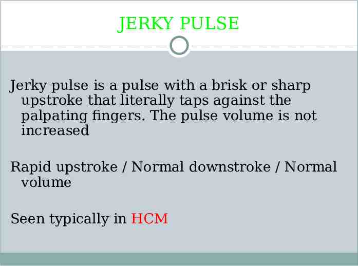
JERKY PULSE Jerky pulse is a pulse with a brisk or sharp upstroke that literally taps against the palpating fingers. The pulse volume is not increased Rapid upstroke / Normal downstroke / Normal volume Seen typically in HCM
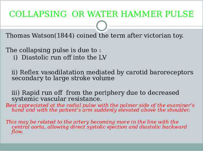
COLLAPSING OR WATER HAMMER PULSE Thomas Watson(1844) coined the term after victorian toy. The collapsing pulse is due to : i) Diastolic run off into the LV ii) Reflex vasodilatation mediated by carotid baroreceptors secondary to large stroke volume iii) Rapid run off from the periphery due to decreased systemic vascular resistance. Best appreciated at the radial pulse with the palmer side of the examiner’s hand and with the patient’s arm suddenly elevated above the shoulder. This may be related to the artery becoming more in the line with the central aorta, allowing direct systolic ejection and diastolic backward flow.
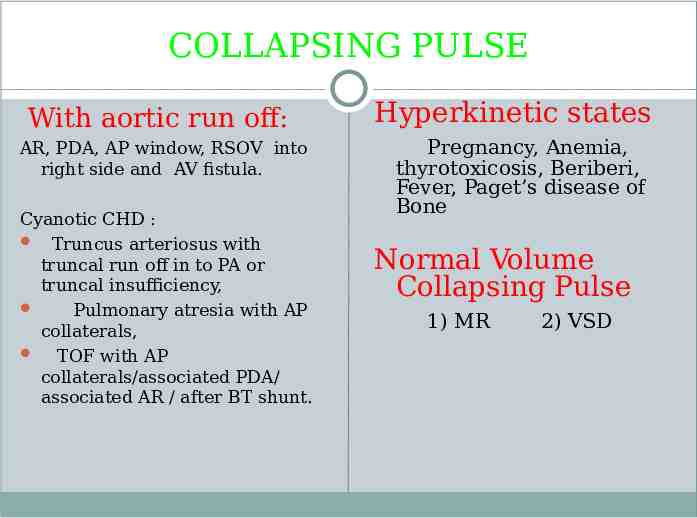
COLLAPSING PULSE With aortic run off: AR, PDA, AP window, RSOV into right side and AV fistula. Cyanotic CHD : Truncus arteriosus with truncal run off in to PA or truncal insufficiency, Pulmonary atresia with AP collaterals, TOF with AP collaterals/associated PDA/ associated AR / after BT shunt. Hyperkinetic states Pregnancy, Anemia, thyrotoxicosis, Beriberi, Fever, Paget’s disease of Bone Normal Volume Collapsing Pulse 1) MR 2) VSD
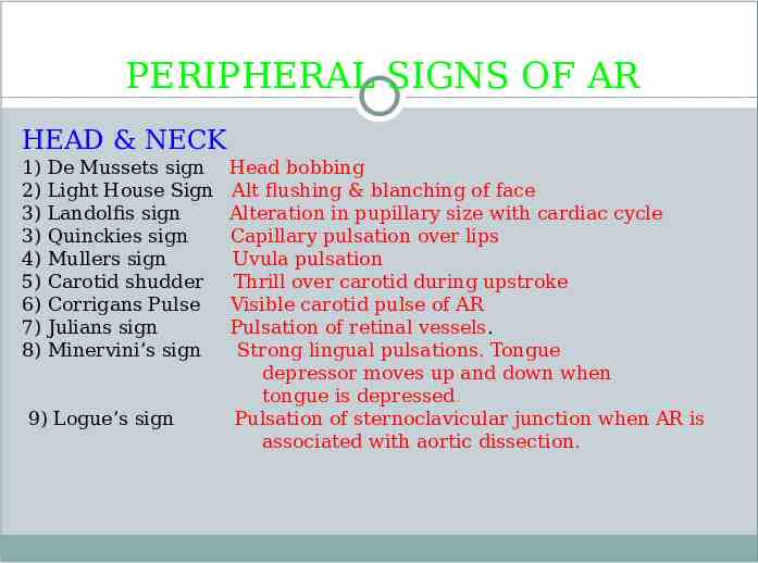
PERIPHERAL SIGNS OF AR HEAD & NECK 1) 2) 3) 3) 4) 5) 6) 7) 8) De Mussets sign Light House Sign Landolfis sign Quinckies sign Mullers sign Carotid shudder Corrigans Pulse Julians sign Minervini’s sign 9) Logue’s sign Head bobbing Alt flushing & blanching of face Alteration in pupillary size with cardiac cycle Capillary pulsation over lips Uvula pulsation Thrill over carotid during upstroke Visible carotid pulse of AR Pulsation of retinal vessels. Strong lingual pulsations. Tongue depressor moves up and down when tongue is depressed. Pulsation of sternoclavicular junction when AR is associated with aortic dissection.
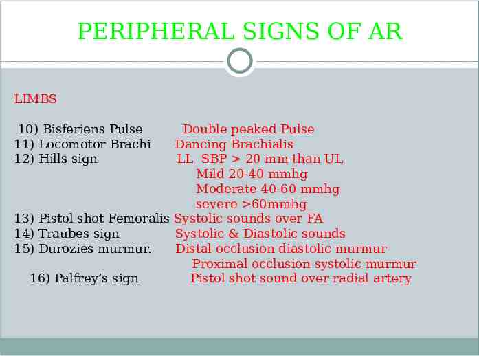
PERIPHERAL SIGNS OF AR LIMBS 10) Bisferiens Pulse 11) Locomotor Brachi 12) Hills sign Double peaked Pulse Dancing Brachialis LL SBP 20 mm than UL Mild 20-40 mmhg Moderate 40-60 mmhg severe 60mmhg 13) Pistol shot Femoralis Systolic sounds over FA 14) Traubes sign Systolic & Diastolic sounds 15) Durozies murmur. Distal occlusion diastolic murmur Proximal occlusion systolic murmur 16) Palfrey’s sign Pistol shot sound over radial artery
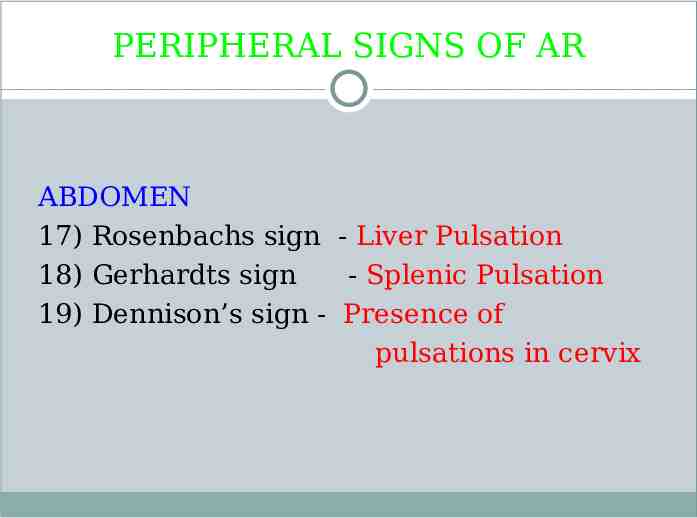
PERIPHERAL SIGNS OF AR ABDOMEN 17) Rosenbachs sign - Liver Pulsation 18) Gerhardts sign - Splenic Pulsation 19) Dennison’s sign - Presence of pulsations in cervix
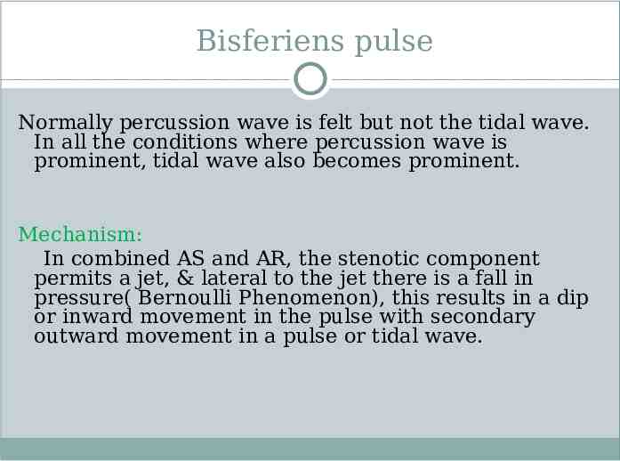
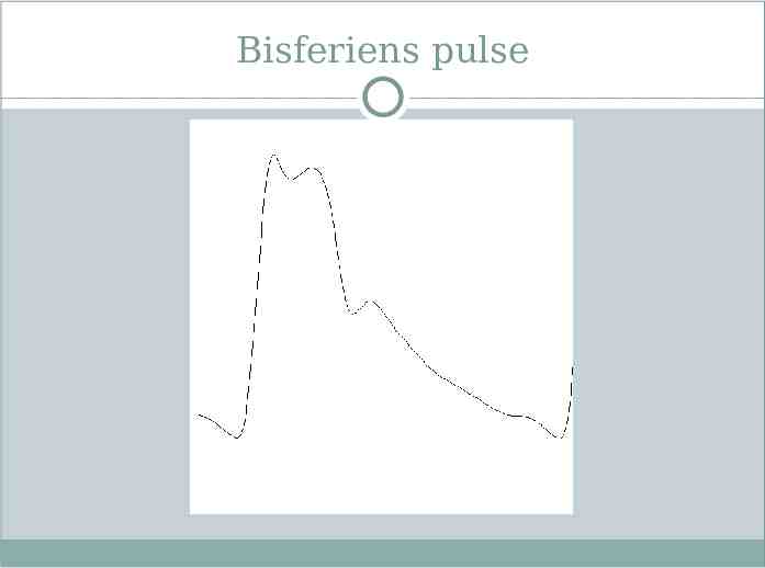
Bisferiens pulse Normally percussion wave is felt but not the tidal wave. In all the conditions where percussion wave is prominent, tidal wave also becomes prominent. Mechanism: In combined AS and AR, the stenotic component permits a jet, & lateral to the jet there is a fall in pressure( Bernoulli Phenomenon), this results in a dip or inward movement in the pulse with secondary outward movement in a pulse or tidal wave.
Bisferiens pulse
Bisferiens pulse Normally both waves are prominent in patients with severe AR. In HOCM, the initial part of left ventricular ejection is rapid, resulting in rapid upstroke. As obstruction to the outflow starts later in the systole, due to SAM, a sudden interruption to left ventricular ejection occurs resulting in a dip in the pressure pulse followed by the slow rising pulse wave, which is characteristic of HOCM ( spike and dome pattern). The percussion wave is more prominent than tidal wave in HOCM. Seen in Severe AR,AS with AR,HOCM,hyperkinetic circulatory state,after exercise
DICROTIC PULSE Dicrotic pulse has an accentuated dicrotic wave and hence is a twice beating pulse, one in systole and one in diastole. Requirements : 1) Hypotension 2) Reduced Peripheral Vascular Resistance When the reflection wave travels rapidly and meets the original wave well in advance, it is lost in it. In rigid and nondistensible arterial system, as in SHT, dicrotic pulse in never present. It is differentiated from the bisferiens pulse by the simultaneous auscultation of the heart sounds.
DICROTIC PULSE It is more noticeable in the beat following a PVC. It is better appreciated during inspiration or inhalation of amyl nitrite. IABP-augmented wave due to diastolic flow occlusion in descending aorta Rarely present when BP 130 mmHg and in patients beyond 50 years of age.
DICROTIC PULSE 1) 2) 3) 4) 5) 6) 7) 8) Healthy young adults Fever Hypovolemic shock CCF Cardiac tamponade Sepsis Post AVR IABP
TWICE BEATING PULSE Anacrotic, Bisferiens ,Dicrotic Differentiation: The double peaking occurs A) On the upstroke in Anacrotic; late peaking B) On the peak in Bisferiens- Both in Systole; rapid rising C) On the downstroke in Dicrotic ; normal rising One in Systole & One in Diastole
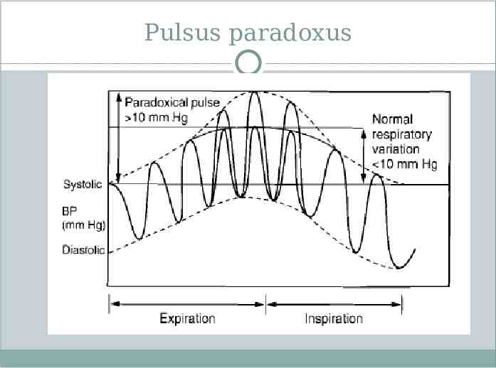
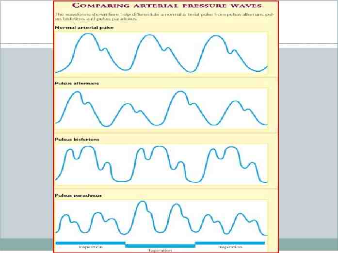
PULSUS PARADOXUS Paradox about the pulse is absence of pulse during inspiration but presence of heart sounds & was coined by Adolph Kussmaul in 1873. Suspected if the pulse varies with inspiration in all accessible arteries. the term paradoxus is that normally there is a fall in BP during inspiration (46mm/hg) which in PP is exaggerated ( 10mm/hg) MISNOMER-
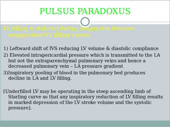
PULSUS PARADOXUS LV filling is reduced during inspiration because exaggerated RV filling causes 1) Leftward shift of IVS reducing LV volume & diastolic compliance 2) Elevated intrapericardial pressure which is transmitted to the LA but not the extraparenchymal pulmonary veins and hence a decreased pulmonary vein – LA pressure gradient 3)Inspiratory pooling of blood in the pulmonary bed produces decline in LA and LV filling. [Underfilled LV may be operating in the steep ascending limb of Starling curve so that any inspiratory reduction of LV filling results in marked depression of the LV stroke volume and the systolic pressure].
Pulsus paradoxus
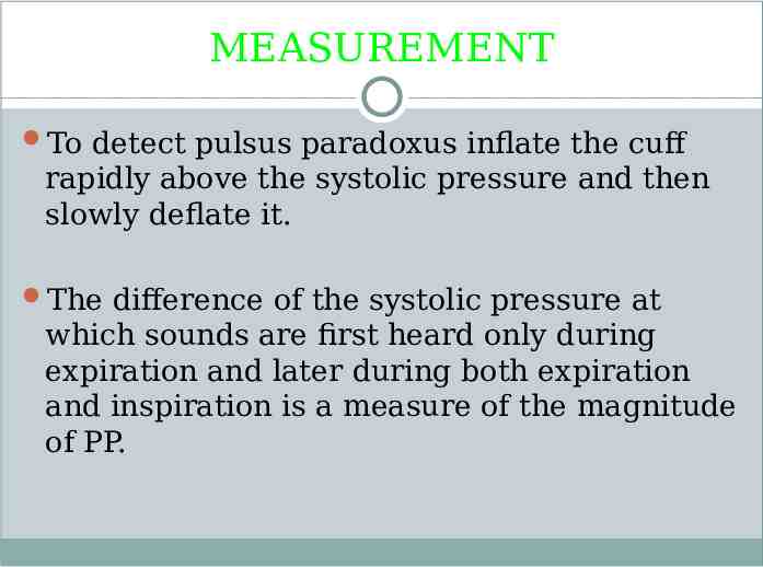
MEASUREMENT To detect pulsus paradoxus inflate the cuff rapidly above the systolic pressure and then slowly deflate it. The difference of the systolic pressure at which sounds are first heard only during expiration and later during both expiration and inspiration is a measure of the magnitude of PP.
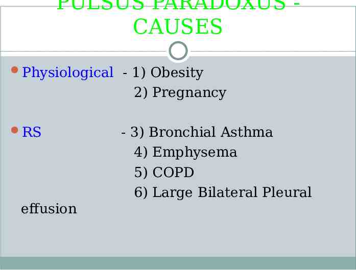
PULSUS PARADOXUS CAUSES Physiological - 1) Obesity 2) Pregnancy RS effusion - 3) Bronchial Asthma 4) Emphysema 5) COPD 6) Large Bilateral Pleural
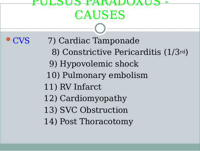
PULSUS PARADOXUS CAUSES CVS 7) Cardiac Tamponade 8) Constrictive Pericarditis (1/3rd) 9) Hypovolemic shock 10) Pulmonary embolism 11) RV Infarct 12) Cardiomyopathy 13) SVC Obstruction 14) Post Thoracotomy
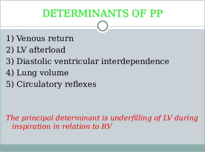
DETERMINANTS OF PP 1) 2) 3) 4) 5) Venous return LV afterload Diastolic ventricular interdependence Lung volume Circulatory reflexes The principal determinant is underfilling of LV during inspiration in relation to RV
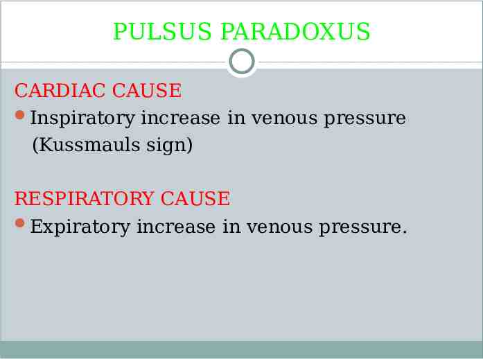
PULSUS PARADOXUS CARDIAC CAUSE Inspiratory increase in venous pressure (Kussmauls sign) RESPIRATORY CAUSE Expiratory increase in venous pressure.
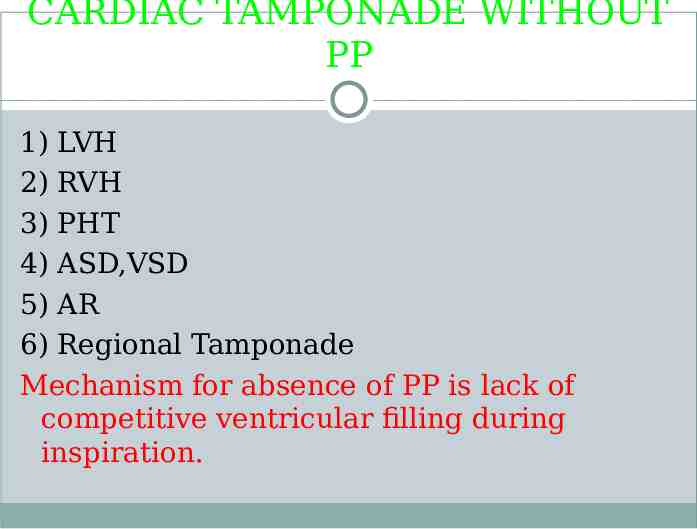
CARDIAC TAMPONADE WITHOUT PP 1) LVH 2) RVH 3) PHT 4) ASD,VSD 5) AR 6) Regional Tamponade Mechanism for absence of PP is lack of competitive ventricular filling during inspiration.
REVERSED PP In Reversed Pulsus Paradoxus there is an increase in systemic pressure with inspiration 1) HOCM : Mechanism unknown. 2) Isorhythmic AV dissociation : Atrial activity precedes QRS during inspiration and marches into QRS during expiration. The atrial activity during inspiration increases the stroke volume and its lack during expiration decreases the stroke volume and systolic pressure. 3) IPPV : Intrathoracic pressure is higher during inspiration and lower during expiration.
PULSUS ALTERNANS Beats occur at regular intervals but in which there is a regular attenuation of the systolic height of the pressure pulse. It was first described by Traube in 1872. Pulsus Alternans is a peripheral manifestation of LV failure 1) Alteration in the height of the pressure pulse 2) Alteration in the rate of rise. It is the latter that is appreciated during palpation.
PULSUS ALTERNANS PA is better felt in distal vessels than proximal- rate of rise & peak pressure developed are accentuated during peripheral transmission of the arterial pulse pressure. Light pressure is applied to palpate Pulsus alternans. Mild degree of PA is detected by sphygmomanometer. Inflate the BP cuff rapidly above SBP and then deflate slowly until Korotkoffs sounds are audible. At this point beats are heard at one half of the heart rate. When the cuff is deflated further the rate doubles.
PULSUS ALTERNANS MECHANISM It is due to alteration of the contractile state of at least part of the myocardium, caused by failure of electromechanical coupling in some cells during weaker contraction. Alternate more and less number of contractile elements participate in each contraction. Correlates with alteration in intensity f Korotkoff sounds.
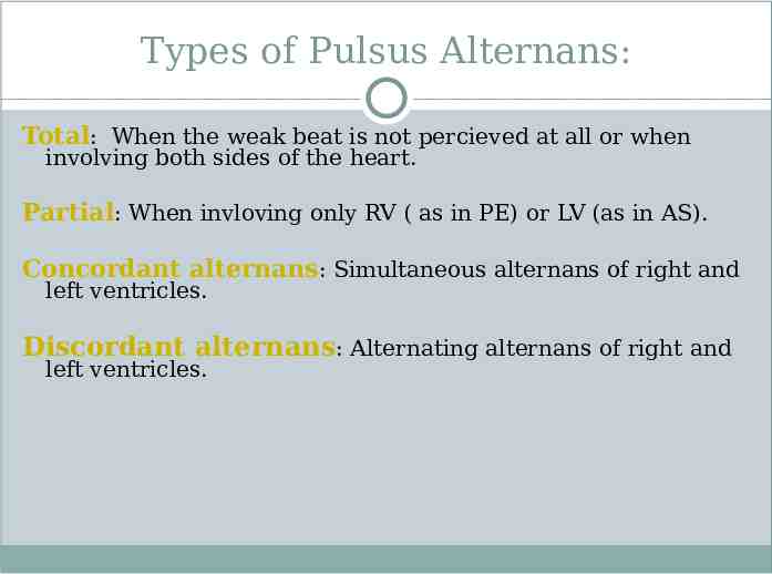
Types of Pulsus Alternans: Total: When the weak beat is not percieved at all or when involving both sides of the heart. Partial: When invloving only RV ( as in PE) or LV (as in AS). Concordant alternans: Simultaneous alternans of right and left ventricles. Discordant alternans: Alternating alternans of right and left ventricles.
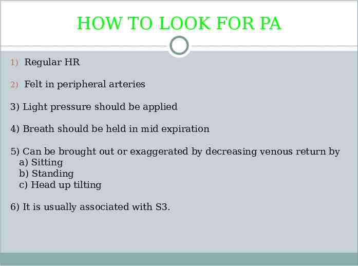
HOW TO LOOK FOR PA 1) Regular HR 2) Felt in peripheral arteries 3) Light pressure should be applied 4) Breath should be held in mid expiration 5) Can be brought out or exaggerated by decreasing venous return by a) Sitting b) Standing c) Head up tilting 6) It is usually associated with S3.
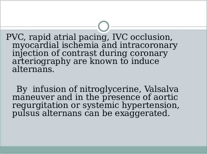
PVC, rapid atrial pacing, IVC occlusion, myocardial ischemia and intracoronary injection of contrast during coronary arteriography are known to induce alternans. By infusion of nitroglycerine, Valsalva maneuver and in the presence of aortic regurgitation or systemic hypertension, pulsus alternans can be exaggerated.
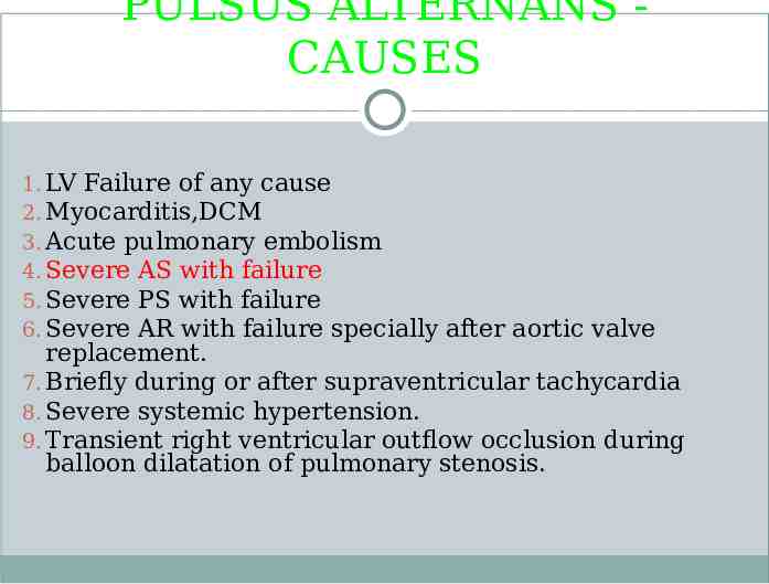
PULSUS ALTERNANS CAUSES 1. LV Failure of any cause 2. Myocarditis,DCM 3. Acute pulmonary embolism 4. Severe AS with failure 5. Severe PS with failure 6. Severe AR with failure specially after aortic valve replacement. 7. Briefly during or after supraventricular tachycardia 8. Severe systemic hypertension. 9. Transient right ventricular outflow occlusion during balloon dilatation of pulmonary stenosis.
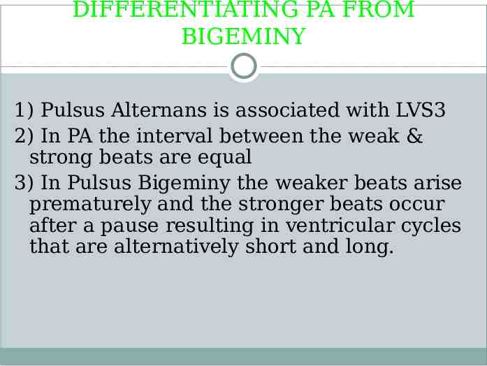
DIFFERENTIATING PA FROM BIGEMINY 1) Pulsus Alternans is associated with LVS3 2) In PA the interval between the weak & strong beats are equal 3) In Pulsus Bigeminy the weaker beats arise prematurely and the stronger beats occur after a pause resulting in ventricular cycles that are alternatively short and long.
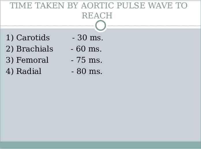
TIME TAKEN BY AORTIC PULSE WAVE TO REACH 1) 2) 3) 4) Carotids Brachials Femoral Radial - 30 ms. - 60 ms. - 75 ms. - 80 ms.
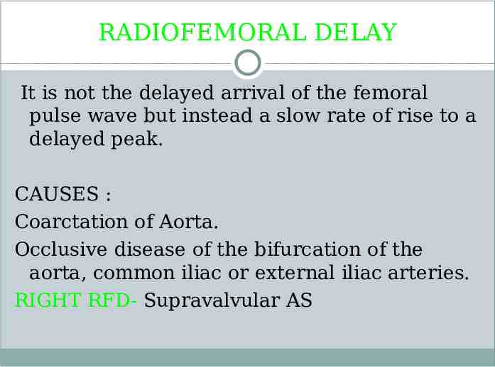
RADIOFEMORAL DELAY It is not the delayed arrival of the femoral pulse wave but instead a slow rate of rise to a delayed peak. CAUSES : Coarctation of Aorta. Occlusive disease of the bifurcation of the aorta, common iliac or external iliac arteries. RIGHT RFD- Supravalvular AS
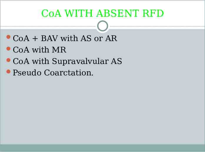
CoA WITH ABSENT RFD CoA BAV with AS or AR CoA with MR CoA with Supravalvular AS Pseudo Coarctation.
PULSE DEFICIT Difference between apex beat and radial pulse 10 beats/mt occurs in AF With VPC if they are too weak to open the aortic valve.
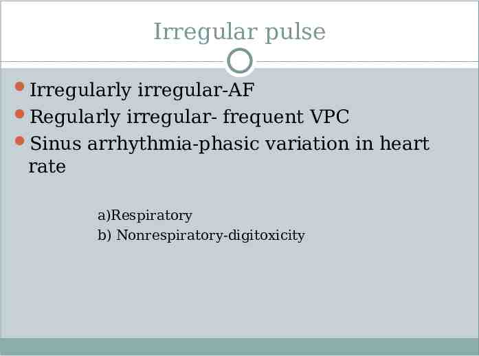
Irregular pulse Irregularly irregular-AF Regularly irregular- frequent VPC Sinus arrhythmia-phasic variation in heart rate a)Respiratory b) Nonrespiratory-digitoxicity
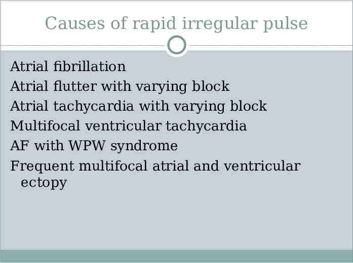
Causes of rapid irregular pulse Atrial fibrillation Atrial flutter with varying block Atrial tachycardia with varying block Multifocal ventricular tachycardia AF with WPW syndrome Frequent multifocal atrial and ventricular ectopy
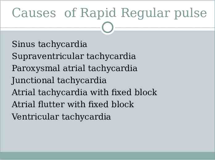
Causes of Rapid Regular pulse Sinus tachycardia Supraventricular tachycardia Paroxysmal atrial tachycardia Junctional tachycardia Atrial tachycardia with fixed block Atrial flutter with fixed block Ventricular tachycardia
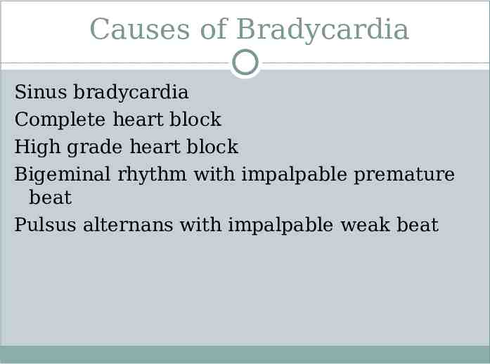
Causes of Bradycardia Sinus bradycardia Complete heart block High grade heart block Bigeminal rhythm with impalpable premature beat Pulsus alternans with impalpable weak beat
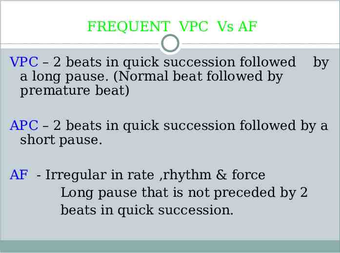
FREQUENT VPC Vs AF VPC – 2 beats in quick succession followed a long pause. (Normal beat followed by premature beat) by APC – 2 beats in quick succession followed by a short pause. AF - Irregular in rate ,rhythm & force Long pause that is not preceded by 2 beats in quick succession.
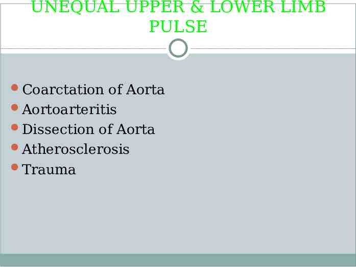
UNEQUAL UPPER & LOWER LIMB PULSE Coarctation of Aorta Aortoarteritis Dissection of Aorta Atherosclerosis Trauma
UNEQUAL CAROTIDS Aortoarteritis Dissecting aneurysm of Aorta Atherosclerosis Thromboembolic occlusion Supravalvular AS
UNEQUAL RADIALS Aortoarteritis Dissecting aneurysm of Aorta Thromboembolic obstruction Previous catheterization Cervical rib Scalenus Anticus syndrome Anomalous Rt Subclavian artery Aberrant course of Radial artery Arteritis.
ABSENT FEMORALS Dissecting aneurysm Coarctation of aorta Pseudoxanthoma elasticum Hypoplastic External Iliac artery.
Points to remember 1)If the arterial pulse is regular in a patient with established atrial fibrillation on digitalis therapy, digitoxicity with AV nodal rhythm should be considered. 2)Presence of dicrotic wave always suggests a grave prognosis. 3) Severe MR with good LV function results in normal volume collapsing pulse. This is due to rapid ejection by the LV with the advantage of lesser afterload and more preload. With the onset of LV dysfunction, pulse loses its collapsing character 4)Electrical alternans has no relationship to pulsus alternans